![]() One of the hallmarks of Autism Spectrum Disorder (ASD) is an impairment in social cognitive skills. This manifests in individuals with ADS having trouble orienting their attention towards people. Accordingly, they also show deficits orienting their attention in response to social cues from others, such as eye gaze, head turns and pointing gestures.
One of the hallmarks of Autism Spectrum Disorder (ASD) is an impairment in social cognitive skills. This manifests in individuals with ADS having trouble orienting their attention towards people. Accordingly, they also show deficits orienting their attention in response to social cues from others, such as eye gaze, head turns and pointing gestures.
Understanding the social cognitive impairments associated with ASD has been challenging in that studies set in naturalistic settings often reveal the deficit but lab experiments performed on computers don’t.
For example, some naturalistic studies have looked at home movies of infants and found that those later diagnosed with ASD showed less social orienting and were less responsive to cues from others to orient to objects. For example, if their mom was in the room, they would look at her a lot less and they’d also be less likely to respond when their mothers tried to direct their attention to a toy in the room by looking or pointing at it.
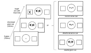
However, people with ASD have been shown to respond to non-naturalistic social cues in the lab. Social orienting has been frequently been tested by use of a variation on Michael Posner’s spatial cueing paradigm. This works as follows:
1. Participants are seated in front of a computer
2. A stimulus – a pair of eyes gazing to either side (or straight ahead) or arrows pointing to either side or neither – appears on the screen
3. Shortly after, a stimulus (the target object) appears to one side or the other, either on the side which the eyes or arrows were pointing towards or the opposite side.
4. Participants have to indicate which side the target object appeared on by pressing either a right or left button.
5. Performance on the task is assessed by measuring the amount of time it takes to participants to press the button indicating on which side the target appeared. Most participants, including ASD patients, are as quick with the gaze cue (the eyes) as with the arrow cue.
(The left side of the above figure shows a single trial (with “directional eyes”), in which participants first see a fixation cross, then one of four directional/non-directional stimuli, after which the target appears either on the same side indicated by the cue or the opposite side. Participants need to indicate which side a target stimulus appeared on by pushing a button. The right side shows the three other trial types (from top to bottom): neutral arrow, directional arrow, neutral eyes)
Past studies have shown that people orient faster to cued (like in the left side of the above figure) versus noncued locations, known as the facilitation effect. Previous studies using this task have produced inconsistent results, but most of them have shown ASD populations performing comparably to non-ASD populations.
In this study, researchers used the above-described cue task to examine the neural mechanisms underlying social orienting in ASD, with the hope that if there were no behavioral differences, neural activity might reveal that ASD individuals are performing the task differently. Other studies have shown that non-ASD populations treat social and non-social cue stimuli differently. It was hoped that neural activity revealed in this study would shed light on the discrepancies in behavioral results for ASD populations in lab versus computer settings.
Results
In terms of behavior, both the control and the ASD group showed quicker responses for gaze and arrow cues with no between group difference, which is consistent with previous lab studies.
However, neural activation patterns showed significant group differences. The control group showed greater activation for social vs. nonsocial cues in many different brain regions, with gaze (eyeball) cues eliciting increased activity in many frontoparietal areas, supporting the idea that neurotypical brains treat social stimuli different from non-social stimuli. The ASD group, on the other hand, showed much less difference in neural activation between social vs. non-social cues. Although these differences in neural activation are too numerous to cover here, one region of interest, superior temporal sulcus (STS), stood out. The STS has been shown to be associated with the perception of eye gaze and other work has suggested the region may be involved in understanding the intentions and mental states of others. In this study, ASD individuals showed decreased STS in the gaze cue condition (versus controls). This data suggests that the STS may not be sensitive toward the social significance of eye gaze in ASD individuals.
Implications
The authors point out that although ASD individuals don’t seem to rely on the same neural circuitry to perceive social cues such as eye gaze, they have found a way to use the low-level perceptual information available in social cues to adapt a strategy that allows them to discern that gaze direction conveys meaning about the environment. That being said, ASD individuals mostly don’t do this very well in more naturalistic environments. So, although this strategy might work in a scanner with “cartoon” eyes and where there are no environmental distractions, it’s unlikely that ASD individuals could adapt this strategy in a naturalistic environment. On the contrary, one could also frame these results from the perspective of the ASD individual; that is, given the non-naturalistic environment of the scanner, and the fact that the task demands were very simple and not dependent on social cognitive processing, why should non-ASD individuals treat the gaze vs. arrow stimuli differently? Why not just rely on low-level information and thus expend less cognitive energy? It’s a good example of the automaticity of social cognitive processes. Give humans a set of cartoon eyeballs to look at and they can’t help but process these as distinct from something non-social.
An additional take away from this paper is that even when one finds no behavioral differences between groups, there might be some interesting differences in neural activity worth exploring via fMRI or EEG.
References
Greene DJ, Colich N, Iacoboni M, Zaidel E, Bookheimer SY, & Dapretto M (2011). Atypical neural networks for social orienting in autism spectrum disorders. NeuroImage, 56 (1), 354-62 PMID: 21334443


You must be logged in to post a comment.