The Neural Correlates of Romantic Love


ResearchBlogging.org

For the most part, fMRI studies attempt to localize cognitive processes to specific regions in the brain. Popular media often introduce these studies with headlines that tout the discovery of “the brain region” for memory, language, empathy, moral reasoning, loving weiner schnitzel and so on.

These headlines can be terribly misleading, as they’re often misinterpreted to suggest a specific brain region is dedicated to a single function, when, in fact, any given function maps on to a network of regions (forming a circuit), while any given region is part of multiple circuits subserving many functions. Similar faux pas can be found in descriptions of the functions associated with genes, e.g. “The gene for (fill in the blank).”

A few years back, the NY Times ran an infamous piece featuring the work of a neuromarketing company. In a horrible experiment fit for The Onion, participants lay in the scanner while looking at pictures of then presidential candidates. Subjects showed increased amygdala activation to pictures of Mitt Romney, which researchers interpreted as a sign of anxiety.

But after watching Romney speak on video, the amygdala activity died down, which researchers said showed that voters’ anxiety had decreased.

Meanwhile subjects’ anterior cingulates lit up to pictures of Hillary Clinton.

Here’s how researchers interpreted this neural activity:

Emotions about Hillary Clinton are mixed. Voters who rated Mrs. Clinton unfavorably on their questionnaire appeared not entirely comfortable with their assessment. When viewing images of her, these voters exhibited significant activity in the anterior cingulate cortex, an emotional center of the brain that is aroused when a person feels compelled to act in two different ways but must choose one. It looked as if they were battling unacknowledged impulses to like Mrs. Clinton.

The Times article about the “research” was quickly and roundly criticized by prominent neuroscientists, 17 of whom quickly responded with a signed letter to the editor, which the Times ran a couple of days later:

To the Editor:

“This Is Your Brain on Politics” (Op-Ed, Nov. 11) used the results of a brain imaging study to draw conclusions about the current state of the American electorate. The article claimed that it is possible to directly read the minds of potential voters by looking at their brain activity while they viewed presidential candidates.

For example, activity in the amygdala in response to viewing one candidate was argued to reflect “anxiety” about the candidate, whereas activity in other areas was argued to indicate “feeling connected.” While such reasoning appears compelling on its face, it is scientifically unfounded.

As cognitive neuroscientists who use the same brain imaging technology, we know that it is not possible to definitively determine whether a person is anxious or feeling connected simply by looking at activity in a particular brain region. This is so because brain regions are typically engaged by many mental states, and thus a one-to-one mapping between a brain region and a mental state is not possible.As cognitive neuroscientists, we are very excited about the potential use of brain imaging techniques to better understand the psychology of political decisions. But we are distressed by the publication of research in the press that has not undergone peer review, and that uses flawed reasoning to draw unfounded conclusions about topics as important as the presidential election.

Adam Aron, Ph.D., University of California, San Diego
David Badre, Ph.D., Brown University
Matthew Brett, M.D., University of Cambridge
John Cacioppo, Ph.D., University of Chicago
Chris Chambers, Ph.D., University College London
Roshan Cools, Ph.D., Radboud University, Netherlands
Steve Engel, Ph.D., University of Minnesota
Mark D’Esposito, M.D., University of California, Berkeley
Chris Frith, Ph.D., University College London
Eddie Harmon-Jones, Ph.D., Texas A&M University
John Jonides, Ph.D., University of Michigan
Brian Knutson, Ph.D., Stanford University
Liz Phelps, Ph.D., New York University
Russell Poldrack, Ph.D., University of California, Los Angeles
Tor Wager, Ph.D., Columbia University
Anthony Wagner, Ph.D., Stanford University
Piotr Winkielman, Ph.D., University of California, San Diego

Undoubtedly, fewer people saw that letter than saw the original article, which was much more prominently displayed.

(By the above study’s logic, looking at a picture of Donald Trump should elicit activity in the anterior insula, a region often associated with disgust responses)

It’s unfortunate that the study received such a prominent platform for distribution because people, especially non scientists, can be heavily influenced by articles with pictures of brains or technical sounding neuro language. One study, which I’ve written about on the blog, found that people were much more likely to believe a nonsensical article if it had meaningless neuroscience language in it than if it didn’t. As the average lay person doesn’t possess the technical skills to distinguish between valid and invalid fMRI studies, it’s up to the scientific community to police itself, which it does a pretty good job of through the peer review process.

This next study I’ll talk about demonstrates some of the challenges inherent to mapping localized neural activity onto unseen mental processes; in this case, the subjective experience of intense, romantic long-term love.

Aaron and colleagues previously published a study that presented neural correlates of intense romantic love (2005). In brief, the study reported that regions in the reward circuit of the brain were activated in response to pictures of a lover (versus a close friend). In the current study, they wanted to explore if these findings could be extended to long-term married couples (couples together for more than 20+ years who report still being madly in love).

Participants lay in the scanner and were repeatedly presented with pictures from four different categories: their partner, a close friend, and both a highly familiar and low-familiar neutral acquaintance. They were instructed to think about “experiences with each stimulus person (that were) nonsexual in nature.”

The fMRI data was analyzed via subtraction, a common fMRI analysis method in which one condition is compared to another to see if any differences fall out. The contrast of interest was between the partner and the close friend. In a cognitive process sense, the only difference thought to exist between perceiving these two individuals was thought to be the subject’s romantic love for one and not for the other. So if neural activity in the close friend condition is subtracted from activity in the partner condition, whatever is left over should represent the neural substrate of romantic love.

Researchers found activation in the ventral tegmental area, substantia nigra and nucleus accumbens (and the hippocampus, which corresponded with reported sex frequency, but the effect seems to be driven largely by two outliers, one of whom reported they have sex almost every day).

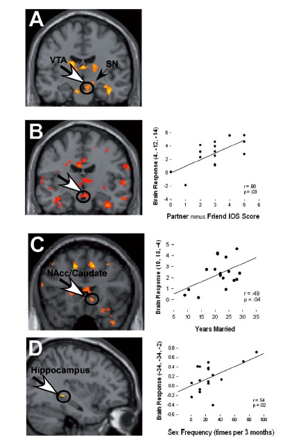
The activity does suggest a classic reward response and replicates previous findings. However, the big question isn’t whether there is a response, but rather what’s driving it?

A valid fMRI study doesn’t only rely on the integrity or analysis of the fMRI data, but, also, and perhaps more importantly, on the experimental design. In order to attribute increased activation in one condition versus the other to a specific cognitive function, one must be confident they have created conditions that have cleanly isolated the independent variable of interest (romantic love). The ventral tegmental area and other regions in the basal ganglia have been repeatedly shown to encode reward value – that is, they respond to things that give us hedonic pleasure, such as food, drugs, sex or receiving money. Past work has shown that intense romantic love is associated with activity in the those regions (Aaron 2005). In the current study, activity in some neural regions previously associated with maternal pair bonding was shown (substantia nigra, for one). The authors hypothesized that neural correlates for romantic long-term love should encompass those associated with both intense romantic love and maternal pair bonding.

But this analysis is dependent on long-term, romantic love being the only difference between conditions that would explain the differences in brain activity. And that may not entirely be the case.

Alternative Explanations
Just to refresh, the major dependent measure of interest was neural activity, especially of reward circuitry, while subjects looked at pictures of their long-term partner versus a close friend. One additional difference between these conditions (beyond romantic love) is that romantic partners are probably more familiar and closer to participants than close friends. This is a shortcoming acknowledged by the authors.

This difference suggests a causal chain of cognitive operations that could offer alternative explanations for some of the data seen in this study. First, It’s been shown that we prefer things that are familiar to us (the “mere exposure” effect, Zajonc, 1968). Second, we’re able to process familiar things (or people) much more fluently compared to the less familiar (Reber). Third, fluency processing has been associated with judgments of aesthetic appreciation such that the more fluently we can process something, the more beautiful or attractive we’re likely to rate it (Alter). Although the objective attractiveness of the photos was controlled for via a group of independent raters, the participants were likely much more subjective in their judgments and perhaps found their partners more attractive than an objective viewer might. Viewing attractive faces has been shown to elicit strong neural activity, particularly in the reward circuitry (NaCC and OFC).

Furthermore, it has been posited that people incorporate close others into their psychological construct of self. Recent studies (deGreck 2008) showed that regions active in a reward task, such as the bilateral ventral striatum, and the ventral tegmental area (VTA), are also involved in differentiating between high and low personal relevance.It seems that we find thinking about ourselves pretty damn rewarding! (We’re all at least a little bit narcissistic). To the extent that someone has been incorporated into our self concept, thinking about that person, or looking at their picture as in this study, could be correlated with responses in reward regions of the brain in part because they activate thoughts of ourselves.

Both the familiarity –> processing fluency –> attractiveness model and self relevant thinking are plausible alternative explanations for at least some of the neural correlates found in this paper.

One other potential area of concern is that there is no way of knowing that participants weren’t thinking about sex with their partners, even though they were told not to. This might be especially difficult to achieve, especially for the two outliers who reported almost daily sex. Regions active during sexual arousal include R. amygdala, hypothalamus, hippocampus, midbrain, mOFC and nucleus accumbens, many of which were found to be active in this study.

The neural activity measured here may very well reflect some aspect of individuals’ love for their partners. But there seem to be other possible explanations for some of the data. I suppose that’s why people call the study of consciousness, of which subjective experiences such as romantic love are a subset, the “hard problem.”

References
Acevedo BP, Aron A, Fisher HE, & Brown LL (2011). Neural correlates of long-term intense romantic love. Social cognitive and affective neuroscience PMID: 21208991

Aron, A., Fisher, H., Mashek, D., Strong, G., Li, H., Brown, L. (2005). Reward, motivation and emotion systems associated with early-stage intense romantic love. Journal of Neurophysiology, 93, 327–37.

DEGRECK, M., ROTTE, M., PAUS, R., MORITZ, D., THIEMANN, R., PROESCH, U., BRUER, U., MOERTH, S., TEMPELMANN, C., & BOGERTS, B. (2008). Is our self based on reward? Self-relatedness recruits neural activity in the reward system NeuroImage, 39 (4), 2066-2075 DOI: 10.1016/j.neuroimage.2007.11.006

Alter, A., & Oppenheimer, D. (2008). Easy on the mind, easy on the wallet: The roles of familiarity and processing fluency in valuation judgments Psychonomic Bulletin & Review, 15 (5), 985-990 DOI: 10.3758/PBR.15.5.985

Peskin, M., & Newell, F. (2004). Familiarity breeds attraction: Effects of exposure on the attractiveness of typical and distinctive faces Perception, 33 (2), 147-157 DOI: 10.1068/p5028

Reber, R., Schwarz, N., & Winkielman, P. (2004). Processing Fluency and Aesthetic Pleasure: Is Beauty in the Perceiver’s Processing Experience? Personality and Social Psychology Review, 8 (4), 364-382 DOI: 10.1207/s15327957pspr0804_3

Unconscious priming studies (for adults only)

The last few years have seen a calvacade of studies demonstrating that unexpected elements in the environment can unconsciously prime attitudes, beliefs and behaviors.

Just a few recent examples:

1. People are more likely to judge a person as “warmer” just after holding a warm (compared to a cold) cup (Williams & Bargh 2008a):
2. Job candidates whose resumes were seen on a heavy (versus light) clipboard were rated as better qualified for a job (Ackerman 2010)
3. The hotter the temperature is in a room or outside, the more likely people are to believe in global warming (Li 2011)
4. Working on a jigsaw puzzle with rough (versus smooth) pieces made people rate a subsequent personal encounter as “less smooth” (Ackerman 2010)

Based on the abundance of findings, there seems to be a robust market in unconscious priming.

Why do we see these kinds of effects? Yale psychologist and implicit cognition guru John Bargh said “… these (kinds of) demonstrations suggest a cognitive architecture in which social psychological concepts metaphorically related to physical-sensory concepts … are grounded in those physical concepts, such that activation of the physical version also activates (primes) the more abstract psychological concept.” What he’s describing is essentially conceptual metaphor theory, which originated with George Lakoff and Mark Johnson in the early 1980s. The theory suggests that cognition is largely based on metaphoric thinking, whereby the structure and logical protocols of one domain guides or structures thinking in another.

Aside from their theoretical contributions, these kinds of studies also provide a certain entertainment value in that they demonstrate associations between a given stimuli and an attitude or behavior that confounds our expectations.

A new study from University of New Mexico researchers presents yet another priming effect that can be added to the ever growing list. But this one ain’t for the kiddies.

In a nutshell: The study showed that the smell of poop, er, uh, fecal matter, (delivered in the form of a spray from a bottle of “Liquid Ass,” a novelty odor liquid) made participants more likely to report their intentions to use condoms in the near future. In other words, the smell of feces motivated safe sex attitudes. Although unpleasant to describe and probably even more so to have carried out, it does fit theoretically with a literature showing that pathogen avoidance/visceral disgust response can function as a mechanism to elicit unexpected effects on seemingly unrelated behaviors and attitudes. I recently discussed a study that looked at the flip side of the same coin. In this study, priming concerns about cleanliness made participants more likely to condemn a slightly immoral sex act and more likely to report conservative political attitudes (Subjects in the experimental condition were standing next to a soap dispenser).

While credit certainly has to be given for being able to work “Liquid Ass” into an experiment, I’m not sure that the researchers have shown pathogen-avoidance concern is the mechanism motivating subjects’ change in attitude.

For one, sufficient control questionnaires don’t seem to have been employed, asking participants to report attitudes in domains other than those in which pathogen avoidance plays a role. Perhaps the nasty scent mediated a more general shift in risk taking or impulsivity. Furthermore, self report is notoriously unreliable at predicting behavior; that is, participants’ answers might in part reflect self presentation concerns as much as shifts in attitude that would result in behavioral change. Finally, it’s possible that the general unpleasantness of the stimuli caused the shift in attitude and not it’s viscerally disgusting nature. One possible control could be an additional condition with an unpleasant, non-pathogen avoidance related stimuli from a different domain (e.g., an unpleasant noise).

Although it would be interesting to see if the effects would still hold up to these slight modifications, I don’t think I would want to be the one to run it…

References
Anderson ML (2010). Neural reuse: a fundamental organizational principle of the brain. The Behavioral and brain sciences, 33 (4) PMID: 20964882

Tybur JM, Bryan AD, Magnan RE, & Hooper AE (2011). Smells like safe sex: olfactory pathogen primes increase intentions to use condoms. Psychological science : a journal of the American Psychological Society / APS, 22 (4), 478-80 PMID: 21350181

Ackerman, J., Nocera, C., & Bargh, J. (2010). Incidental Haptic Sensations Influence Social Judgments and Decisions Science, 328 (5986), 1712-1715 DOI: 10.1126/science.1189993

Li Y, Johnson EJ, & Zaval L (2011). Local warming: daily temperature change influences belief in global warming. Psychological science : a journal of the American Psychological Society / APS, 22 (4), 454-9 PMID: 21372325

ResearchBlogging.org

The case of the man who couldn’t find the beat

This post was chosen as an Editor's Selection for ResearchBlogging.org

The ability to dance to music comes naturally to most members of the human species, and even exists in some species of bird, most famously a cockatoo and YouTube celebrity named Snowball.

But it doesn’t come naturally to everyone.

Researchers from McGill University and the University of Montreal (Phillips-Silver, 2011) have recently published a case study of a student named Matthieu, who not only can’t dance to the beat, but also can’t tell when someone else is dancing asyncronously, although he can dance in time if he is able to watch someone else doing it.

“Mathieu was discovered through a recruitment of subjects
who felt they could not keep the beat in music, such as in clapping
in time at a concert or dancing in a club. Mathieu was the
only clear-cut case among volunteers who reported these problems.
Despite a lifelong love of music and dancing, and musical
training including lessons over several years in various instruments,
voice, dance and choreography, Mathieu complained that
he was unable to find the beat in music. Participation in music
and dance activities, while pleasurable, had been difficult for
him.”

Experimenters put Matthieu and a group of control subjects through a series of tests in which they danced to various types of music. Measurements were gathered by way of a Wii controller (which contains a accelerometer) that was strapped to the trunk of each subject’s body and was able to track and quantify their movements. They also had participants tap their hands to the beat, while not dancing. Finally, they watched videos of someone else dancing (increasingly out of sync) to some Merengue music, and were to asked to identify if the person dancing in the videos was in sync with the music or not.

See the videos here.

Matthieu couldn’t tap a beat in time and the style of music didn’t seem to matter; across numerous styles of music, he couldn’t dance in sync with the groove.*

*He was able to sync himself somewhat to a techno beat, which is basically a glorified metronome but nonetheless slightly more complex.

However, he had no problem locking his movements to the beat of a metronome and could bounce with a consistent tempo without music, while showing normal levels of pitch and tonal perception. He demonstrated normal intelligence, presented no history of neurological or psychiatric disorders and showed so signs of obvious cognitive deficits. It seems Matthieu’s deficit is specific to perceiving the underlying pulse in a piece of music and moving his body to it. In other words, he’s got beat (rhythm) deafness.

Scientists have been aware of the condition for quite a while.

In an Australian Medical Journal from 1890, a surgeon from the Victorian Eye and Ear Hospital in Melbourne described a case of rhythm deafness in a 27-yr. old farmer named W.M.:

(Unlike Matthieu, the farmer’s deficit was much less selective; he also suffered from tone deafness and had severely reduced pain sensitivity)

More recently, Oliver Sacks touched upon rhythm blindness in his book Musiciophilia:

Google and PubMed searches find numerous casual references to “rhythm deafness”, but this does seem to be the first well documented case in the scientific literature. So, if its been talked about for so long but documented so infrequently, how rare is it?

Lead author Jessica Phillips-Silver suggested that it might be as rare as tone deafness, which affects about 4 to 5% of the population. If that’s the case, it could be a real challenge locating enough participants to conduct an fMRI study, which would help reveal the neural regions implicated in the condition. But the research team is confident, in part due to ample press coverage of the paper, that they’ll find more subjects.

So, what might an fMRI study reveal about the condition?

A 2005 study (Brown) examining the neural substrates of dance points to one possibility. In this study, subjects lay in a PET scanner and danced a tango with their legs only, both accompanied by music and free form (without music).


Participants in the dancing-to-music condition showed BOLD activation suggesting that audio-motor entrainment might be mediated through a connection between subcortical auditory areas and the cerebellum. This would make sense give that one of the primary functions of the cerebellum is to coordinate motor actions, particularly precision and accurate timing, by receiving input from the sensory system and integrating those incoming signals to execute fine tuned motor activity.

The authors suggest that the deficit might be primarily perceptual and point to the fact that he failed on a task which did not require body movement, nor does not have any basic motor impairments They also suggest that basal ganglia connections between auditory and motor cortices could play a role, particularly the dorsal auditory pathway leading to the dorsal premotor cortex. Silver and colleagues already have some neuroimaging work underway with Matthieu.

As for future directions, Silver-Phillips said that her group will be looking at exactly what level of musical complexity is required for Matthieu’s beat deafness to emerge. They’re also interested in exploring whether there is any sign of entrainment occurring on a neuronal level, even in the face of the behavioral deficit. In other words, maybe his neurons are dancing to the beat even if he’s not.

References
Phillips-Silver J, Toiviainen P, Gosselin N, Piché O, Nozaradan S, Palmer C, & Peretz I (2011). Born to dance but beat deaf: A new form of congenital amusia. Neuropsychologia, 49 (5), 961-9 PMID: 21316375

Brown, S. (2005). The Neural Basis of Human Dance Cerebral Cortex, 16 (8), 1157-1167 DOI: 10.1093/cercor/bhj057

ResearchBlogging.org

Regard thyself and put down the smoke stick

As many a former smoker will probably attest, quitting cigarettes ranks high in the hard-to-kick category. I made several unsuccessful attempts before finally kicking the habit after a 10 year pack-a-day run. Ultimately what worked for me was to go cold turkey, but there were perhaps other alternatives which I might have tried. In a paper from Nature Neuroscience, researchers from University of Michigan provided participants with interventions involving individually tailored messages* designed to encourage quitting and found that participants’ brain activity while listening to the messages predicted how likely they would be to successfully quit smoking.

*Tailored messages are statements about an individuals’ issues and thoughts about quitting smoking, derived from pre-screen interviews with them. e.g., “You are worried that when angry or frustrated, you may light up”.

Here’s the premise: Anti-smoking messages custom made for an individual can be more effective than generic ones, but only if said individual processes those messages in a self directed manner. Past research has shown a specific set of neural regions – primarily the mPFC and precuneus/posterior cingulate – to be associated with self referential thinking. Therefore, researchers hypothesized, activity in these brain regions while processing tailored anti-smoking messages might predict the likelihood of quitting.

The Study
The experiment was carried out over three days with a follow-up visit four months later.

Day 1: 91 participants completed a health assessment, demographic questionnaire and a psychosocial characteristics scale related to quitting smoking. Responses were then used to create smoking cessation messages tailored to each individual.
Day 2: Participants went into scanner and performed 2 fMRI tasks: The first task had participants listen to anti-smoking messages of three different types: personally tailored anti-smoking, non-tailored anti-smoking and neutral.

Here are some examples of what they heard:

Tailored messages
A concern you have is being tempted to smoke when around other smokers.
Something else that you feel will tempt you after you quit is because of a craving.
You are worried that when angry or frustrated, you may light up.
Untailored messages
Some people are tempted to smoke to control their weight or hunger.
Smokers also light up when they need to concentrate.
Certain moods or feelings, places, and things you do can make you want to smoke.
Neutral messages
Oil was formed from the remains of animals and plants that lived millions of years ago.
Sighted in the Pacific Ocean, the world’s tallest sea wave was 112 feet.
Wind is simple air in motion. It is caused by the uneven heating of the earth’s surface by the sun.

Then, participants completed a self appraisal task to identify brain regions active during self relevant thought processes. In this task, participants saw adjectives appear on the screen and had to either rate how much the adjective described them or whether the adjective was positive or negative.

Day 3: Participants completed a web-based smoking cessation program and were instructed to quit smoking. (They were given a supply of nicotine patches to get themselves started)

Results
Behavioral
Experimenters checked in with subjects four months later to see if they were abstaining from smoking. Out of 87 who participated in the smoking cessation program, 45 were not smoking, while 42 were still (or had quit briefly and restarted) smoking.

Subjects were given a surprise memory test for the anti-smoking messages they’d received four months prior and remembered self relevant, tailored messages most well. However, their memory performance was not related to whether they successfully quit smoking.

fMRI
As for the fMRI data, experimenters used a mask of tailored vs. untailored message conditions AND self-appraisal to identify the region common to both processes. This seems like a mild case of double dipping, no? That is, finding a brain region that responds to the condition of interest (in this case, voxels more active in tailored vs. untailored conditions) and then using the same data to test the hypothesis. Ideally, the ROI would be obtained independently of the main task.

A blow by blow on the different contrasts of interest:

1. Researchers looked at brain regions more active during tailored vs. untailored messages and found differential activation in the regions below.

There are, I think, some problems here; mainly, that the task differences for processing tailored vs non-tailored statements may extend beyond self relevant thinking to (1) memory processes employed in processing either category of stimuli; that is, episodic (tailored) vs. semantic (non-tailored), (2) cognitive effort, (3) elicitation of visual vs. non-visual memory, (4) processing fluency and (5) affect or reward responses. Thus, the difference in brain activation found in this task might reflect something other than just self referential processing.

2. The localizer task (used to isolate neural areas involved in self appraisal) had participants process adjectives either by relating them to self or by judging their affective value. This suggests an alternative explanation for the categorical contrast in that it isn’t specific to self per se, but really more specific to people vs. non-people. A more widely used version of this task has participants process adjectives with regards to self or an other. As a further control, a third condition is often included in which participants identify whether words are in upper case or lower case. The contrast applied is (self – control) – (other – control). It’s not clear why the researchers chose the task they did, which seems significantly noisier.

Here’s the contrast from the present study:

And here’s a contrast from another study (Jenkins 2010) that looked at three different types of self-referential processing.

Although roughly similar, the current study shows cortical midline activation seems to be much more dorsal than that found in Jenkins (2010). Using an ROI derived from this localizer task to correlate neural activity in tailored vs. untailored statements with quitting led to a non-significant result (from supplementary materials). This could explain why the researchers used the composite mask to define the ROI.

3. Again, the primary ROI was defined as a composite of overlapping regions between the self reference task AND the tailored vs. untailored statements task, which was used to compare neural activity with quitting behavior. They found that activity in these regions – which included dmPFC, precuneus and angular gyrus – during tailored smoking cessation messages predicted the likelihood of successfully abstaining from smoking. dmPFC and precuneus activation also individually predicted smoking cessation success, although angular gyrus did not.

This study provides clear evidence that participants processed tailored vs. non-tailored messages about smoking differently, and that this difference corresponded to their ability to stop smoking. However,
(1) neither task effectively isolates self referential processing,
(2) the region of activation was much more dorsal than that usually found in this literature (Northoff & Bermpohl, 2004; Schneider et al.,2008; Uddin, Iacoboni, Lange, & Keenan, 2007; Gillihan & Farah, 2005),
(3) an independently obtained ROI yielded insignificant results and
(4) mPFC and precuneus subserve an untold number of cognitive processes beyond self reflection.

Therefore, it seems a bit of a stretch to claim the neural activation found in this study is indicative of self referential processing.

References
Chua HF, Ho SS, Jasinska AJ, Polk TA, Welsh RC, Liberzon I, & Strecher VJ (2011). Self-related neural response to tailored smoking-cessation messages predicts quitting. Nature neuroscience, 14 (4), 426-7 PMID: 21358641

Jenkins AC, & Mitchell JP (2010). Medial prefrontal cortex subserves diverse forms of self-reflection. Social neuroscience, 1-8 PMID: 20711940

Northoff, G. (2005). Emotional-cognitive integration, the self, and cortical midline structures Behavioral and Brain Sciences, 28 (02) DOI: 10.1017/S0140525X05400047

Gillihan, S., & Farah, M. (2005). Is Self Special? A Critical Review of Evidence From Experimental Psychology and Cognitive Neuroscience. Psychological Bulletin, 131 (1), 76-97 DOI: 10.1037/0033-2909.131.1.76

SCHNEIDER, F., BERMPOHL, F., HEINZEL, A., ROTTE, M., WALTER, M., TEMPELMANN, C., WIEBKING, C., DOBROWOLNY, H., HEINZE, H., & NORTHOFF, G. (2008). The resting brain and our self: Self-relatedness modulates resting state neural activity in cortical midline structures Neuroscience, 157 (1), 120-131 DOI: 10.1016/j.neuroscience.2008.08.014

UDDIN, L., IACOBONI, M., LANGE, C., & KEENAN, J. (2007). The self and social cognition: the role of cortical midline structures and mirror neurons Trends in Cognitive Sciences, 11 (4), 153-157 DOI: 10.1016/j.tics.2007.01.001

ResearchBlogging.org

worthy links: Rama, acupuncture, disgust and politics (redux), mouse sex

V.S. Ramachandran gives a stimulating TED lecture and a short interview, mostly about mirror neurons, over at Neurophilosophy.

NeuroLogica’s Steven Novella recaps a recent study showing that acupuncture doesn’t work — and points out the clever way in which the failure was spun by the study’s authors.

Just the other day, I summarized a recent study looking at the relationship between disgust/purity and political ideology. In this short video, a brief discourse by Yale’s Paul Bloom on the topic.

Over at Not Exactly Rocket Science, Ed Wong talks about work out of Bejing that showed male mice with reduced serotonin levels became less choosy about the sex of their sexual partners. And as should be expected, the sensationalistic headlines from mass media organizations shortly followed. From the BBC: “Sexual preference chemical found” And from CBS News, a NY Post-worthy headline: “Serotonin sex bomb: How to make a mouse bisexual or just really horny”